AI Research Engineer Bridging Science & Industry
Specializing in computer vision, scientific machine learning, and research-to-production systems. PhD in AI with proven enterprise deployment experience.

Specializing in computer vision, scientific machine learning, and research-to-production systems. PhD in AI with proven enterprise deployment experience.
Cutting-edge AI research with real-world applications
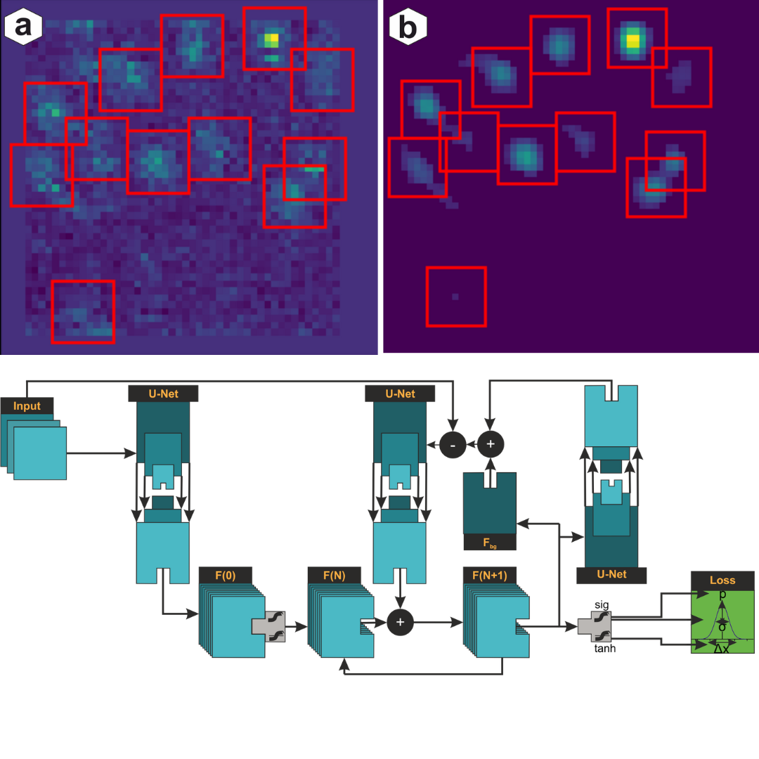
Combined compressed sensing with deep learning for super-resolution microscopy. Achieved state-of-the-art precision for irregular PSFs.
Developed novel algorithms for biological image analysis, registration, and automated quality control with nanometer precision.
SAP consulting with AI showcases. Built production-ready ML systems including EndureXAI platform for sports analytics.
From research prototypes to production systems
Production Sports Analytics Platform
Full-stack AI platform for endurance sports analysis. Features ML-powered performance prediction, training optimization, and real-time analytics.
AI for Scientific Imaging
Revolutionary approach combining compressed sensing with deep learning for confocal lifetime localization microscopy. State-of-the-art precision.
Latest research in AI and computer vision
BMC Bioinformatics, 2022
Novel approach combining deep learning with compressed sensing for super-resolution microscopy...
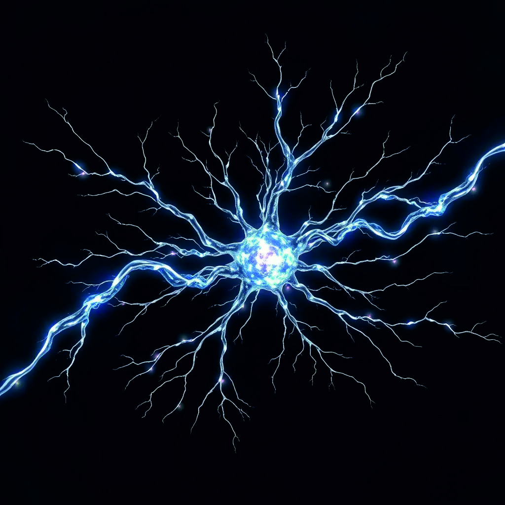
Frontiers in Cellular Neuroscience, 2024
Advanced imaging techniques combined with AI-powered analysis for biological structures...
Let's discuss how AI can solve your most challenging problems